Symptom finder - The causes of breast lump
Symptom finder - The causes of breast lump
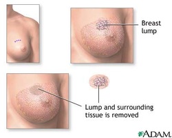
There are many causes of breast lump. Breast lump may affect male and female. Breast lump is a worrying signs of breast carcinoma.
The causes of breast lump are divided into generalized swelling, discrete lesions and swelling located behind the breast. Generalized swelling of the breast is associated with puberty, pregnancy, mastitis and lactation. Malignant disease lumps are due to phyllodes tumor and carcinoma. Breast carcinoma may be associated with lump on the breast and may occur after menopause. Benign discrete lumps are associated with tuberculous abscess ( in TB endemic area), lipoma, cystic mastitis, fat necrosis, galactocele, sebaceous cyst, duct ectasia and fibroadenoma.
Deformities of the rib, Tietze’s disease, chondroma of costal cartilages and chest disease related retromammary abscess are the common causes of swelling behind the breast.
Duct ectasia is presented commonly in the fifth decades as nipple retraction, retro areolar pain and creamy thick nipple discharge. Tuberculous abscess may form in patient with past history of tuberculosis. Tuberculous abscess may erode through the chest wall from the lung or accumulates within the breast and leads to benign discrete lump. Trauma history is associated with duct ectasia. History of cystic hyperplasia , late menopause, early menarche, nulliparous state and family history are associated with breast carcinoma. 85% of breast carcinoma are painless in nature. Patient may notice lump on the breast, dimpling of the skin, nipple retraction and axillary swelling. Headaches, fits, personality changes are suggestive of cerebral metastases. Bone pain occur due to pathological fractures as a result of secondary metastases. Any porta hepatis node involvement or liver metastases may lead to jaundice. Shortness of breath occur due to pleural effusion or secondaries metastases.
Fibroadenoma commonly affect patient between 15 and 25 years of age and present with non tender swelling. Galactocele, mastitis and abscess are associated with pregnancy and lactation.
On examination, galactocele presents as mobile smooth swelling that occur in the lactating breast tissue. Cyst is a mobile, smooth and may be tender lump/swelling. Cyst may present as discrete lump or generalized lump in the breast. Lipoma is uncommon in the breast. It may be difficult to differentiate from pseudolipoma which is a bunching of fat that present between the breast retracted suspensory ligament. Pseudolipoma is related to breast carcinoma. Lipoma is presented as lobulated soft swelling of the breast.
Phyllodes tumor is malignant tumor that present as discrete lump which may become a mobile mass that will become very huge/large. Fat necrosis is difficult to distinguish from breast carcinoma. Fat necrosis is an irregular hard swelling that overlie the teeth marks or bruising of the skin. Fat necrosis will fix to the skin. Fibroadenoma is a rounded mobile smooth mass which may be dart about under the fingers of the examinaer( breast mouse).
Breast carcinoma is an irregular hard mass that fixed superficially or deep to the skin. Commonly present with nipple retraction, skin dimpling and Paget disease of the nipple ( ulcerated eczematous lesions) as well as Peau’d orange. Other signs are hepatomegaly, supraclavicular lymphadenopathy and axillary lymphadenopathy . Long bones, spine and rib are associated with pathological fractures.
There will be tenderness and prominence over the second, third and fourth costal cartilage in cases of Tietze’s syndrome . Chest auscultation and chest percussion may reveal chest signs which are associated with retromammary abscess. Mastitis may present as enlarge, hot, tender red mass. Generalized swelling of the breast is caused by pregnancy, puberty and lactation.
Sebaceous cyst is a mobile cyst with a punctum in certain cases. Sebaceous cyst may be infected and redness of the surrounding and discharge may occur. Duct ectasia presents as tender erythematous retroareolar area with thick creamy, thick nipple discharge and nipple retraction.
The investigations required are full blood count, ESR, liver function test, chest x ray, serum calcium , FNAC, BRCA1, BRCA2, open excision biopsy, mammography, bone scan, CT scan and ultrasound scan.
Full blood count may reveal increase in white cell count due to breast abscess and low hemoglobin level due to malignancy. Raised ESR is associated with tuberculosis or malignanct. There will be a raised in alkaline phosphatase while performing liver function test which indicates the evidence of breast secondaries or liver secondaries / metastases. Chest x ray may reveal the present of empyema or tuberculosis ( lung disorder) or secondary deposition in the rib or lung. Bone secondaries/ metastasis may reveal raised serum calcium.
