|
|
Pathology definition - Urolithiasis

Urolithiasis
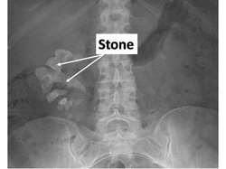
Urolithiasis is also known as kidney stone.The stone usually present may also present at bladder, renal pelvis and renal calyces. Kidney stone can be divided into groups based on the component of the stones. There are calcium phosphate/calcium oxalate stones, cystine stones, uric acid stones and ammonium magnesium phosphate stones.
Calcium phosphate or calcium oxalate stones ( radiopaque appearance on x ray) may occur as a result of sarcoidosis, hyperparathyroidism and intoxication of vitamin D. All of these conditions are associated with hypercalcemic condition. Cystine stone ( radiolucent appearance on x ray) is associate with cystinuria which is a hereditary disorder due to the failure in reabsorption of cystine. Leukemia hyperuricemia and myeloproliferative disorders may lead to uric acid stone. Proteus vulgaris is urease positive bacteria which lead to the formation of ammonium magnesium phosphate stones or struvite stones ( radiopaque appearance on x ray).
Urolithiasis or kidney stones may present with hematuria or pain in the flank region which radiates to the groin. The common complications of urolithiasis are hydronephrosis ( dilation of the renal calyces and renal pelvis which may lead to atrophy of the kidney), pyelonephritis and renal colic. There will also an increase risk of developing urinary tract infection. Patient with kidney stone is advised to increase the intake of fluid and consider surgical removal of the stones. Other treatment may include allopurinol for uric acid stone and hydrochlorothiazide for calcium stones.
References
1.Robertson, W. G., and M. Peacock. "Pathogenesis of urolithiasis." Urolithiasis: Etiology· Diagnosis. Springer Berlin Heidelberg, 1985. 185-334.
2.Polinsky, M. S., B. A. Kaiser, and H. J. Baluarte. "Urolithiasis in childhood." Pediatric Clinics of North America 34.3 (1987): 683-710.
