Paronychia treatment

Paronychia treatment
Paronychia is an acute or chronic condition which affects the fold of the skin surrounding the toenail and fingernails. Paronychia is associated with eczematous or inflammatory condition. Paronychia mostly affect individual who often wet the hands such as nurses, waitresses and bartenders.The acute case of paronychia often present for 2- 5 days after trauma. Paronychia is also known as perionychia or eponychia. It is mostly affecting the endocrine and dermatology system. Children ( infants or toddlers ) are predispose to paronychia due to E.Coli and anaerobes infection cause by thumb /finger sucking.
In term of epidemiology, paronychia is common in united states. It effects women more than men and predominantly affect all ages. The risk factors for developing paronychia may include ingrown nail, trauma o the skin surrounding the nail, diabetes mellitus and sculptured or manicured nail. Anyone with immunosuppression condition (the use of epidermal growth factor inhibitors or antiretroviral therapy for HIV) or frequent immersion of the hand in the water (swimmers, house keepers, bartenders, chefs, cooks).
The general prevention of paronychia may involve good control of diabetics, wear rubber gloves and cloth liner, avoid allergen, keep fingers dry and consider treatment of candidiasis.
Paronychia is caused by infection of the lateral nail fold. The infection may involved complete margin of the skin surrounding the nail plate. This usually occur due to the seperation of the nail plate from the perionychium.
In less than 24 hours, when the infection begin, cellulitis is the initial presentation. If the infection doesn’t resolve an abscess will be formed. In chronic case of paronychia, the paronychia is presented as eczematous reaction with multifocal etiology.
The acute phase of paronychia is caused by infection with streptococcus pyogenes or staphylococcus aureus. Less frequently, It is caused by pseudomonaspyocyonea and proteus vulgaris. Fusobacterium , eikenella corrodens and peptostreptococcus are commonly infected the digits and causing paronychia.
The chronic case of paronychia is associated with eczematous reaction with candida albicans , dermatophytes or mold such as fusarium ir scytalidium. paronychia is commonly associated with diabetes mellitus, eczema or atrophic dermatitis. Certain medication may also trigger paronychia such as paclitaxel, antiretroviral therapy and cetuximab. Multiple paronychia is associated with pemphigus vulgaris.
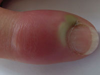
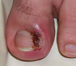
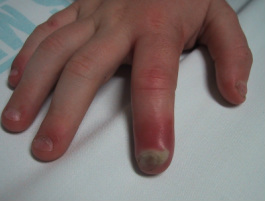
Patient who suffer from paronychia may complain of tenderness and pain which is localized, history of previous trauma such as ingrown nail or bitten nails, contact with herpes infection and contact with irritants and allergens. Physical examination may reveal ( in acute case), hot, red, tense, tender nail fold with or without abscess. Ocassionally the nail bed is elevated and the nail bed is elevated from the nail plate. There will be an area of swollen painful red skin around the nail plate with purulent discharge. There will be a secondary changes of the nail and green color changes in the nail due to pseudomonas infection. The digital pressure test is also positive. The test is performed by applying light pressure over the affected areas and there will be a blanch of the digit and demarcation of the abscess.
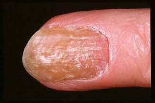
The prominent of chronic paronychia may involve thickening of the nail plate, absence of adjacent cuticle, retraction of the nail fold and the present of Beau’s line ( prominent transverse ridges).
No lab or diagnostic test is required unless the patient suffer from methicillin resistant staphylococcus aureus, resistant to treatment or suffer from severe case of paronychia . The investigation may include culture sensitivity test, gram stain as well as potassium hydroxide wet mount and fungal culture.
The surgical or diagnostic procedure required in paronyhcia may involved scraping for wet mount , incision and drainage for case of suppurative which is non responsive to conservative management. If virak case is suspected consider viral culture and Tzanck testing.
The differential diagnosis of paroncyhia may include Reiter disease, Psoriasis, allergic contact dermatitis ( scrylic, latex), abscess of the pulp of the fingertips (telon) , herpatic whitflow, malignant melanoma, squamous cell carcinoma, metastases, psoriasis eczema and Reiter’s syndrome.
The first line of medication to treat paronychia may involve tetanus booster if appropriate. In acute case which is mild in nature consider antibiotic cream or topical steroid.The antibiotic cream may involve mupirocin, neomycin, gentamicin or polymycin 3 or 4 times a day for 5- 10 days. Betamethasone 0.05 % cream is a topical steroid which is applied twice a day for 7- 14 days.
In acute case as a result of oral exposure consider clindamycin or amoxicillin. Any acute exposure with no oral flora exposure may required dicloxacillin or cephalexin. In acute case when MRSA is suspected consider sulfamethoxazole or trimethoprim or doxycline.
Chronic case of paronychia is treated with betamethasone 0.05% twice daily for 7- 14 days. ( topical steroid ) with or without topical antifungal such as nystatin, clotrimazole or econazole. Three times daily for 30 days. Erythromycin is considered in any case where the patient develop allergies to antibiotics. Precaution should be taken as it may cause gastrointestinal upset. Erythromycin may interact with certain drugs such as antifungal ( ketoconazole, fluconazole, itraconazole,astemizole), terfenadine or statin. Erythromycin may also cause cardiac toxicity due to reaction with astemizole or terfenadine . Theopylline level is also affected by erythromycin.
The second line of treatment may include systemic antifungal therapy such as fluconazole, terbinafine ( lamisil) and itraconazole ( sporanox) . Antipseudomonal therapy such as aminoglycoside and 3rd generation cephalosporin are also considered.
Other additional treatment may include water or ( 1:1) vinegar/ water soak 3 or 4 times daily as well as elevation and warm compression. These measures are useful for acute case. In chronic case, after hand washing, moisturizing lotion is applied, fingers are keep dry, avoid and prevent exposure to irritants and improve diabetic control.
Patient may be refer to the hospital in case of failure of the treatment or recalcitrant where biopsy is considered. Patient with chronic case of paronychia may also be referred for en bloc excision of the nail fold or considering the eponychial marsupilization with or without nail removal. Surgical procedure may involved incision and drainage or partial or complete removal of the nail in case of ingrown nail or subungual abscess.
The patient is advice to avoid any trigerring factors , allergen, frequent immersion , finger sucking or nail bitting. Appropriate diet control is required in diabetic patient and remember to wear rubber gloves with liner of cotton to prevent any exposure to moisture.
Healing is expected with adequate presentation and treatment. Rarely, malignant or benign neoplasm is suspected in any chronic case which is unresponsive to treatment . Patient should be referred. The acute complication of paronychia is subungual abscess while the chronic complication is loss of nail, discoloration, thickening and second ridging of the nail.


