
Emergency Strategy - How to treat volvulus
Emergency Strategy - How to treat volvulus
Patient will be admitted if suspected to suffer from volvulus. Any child who is less than 1 month old and suffer from vomiting should be suspected to suffer from volvulus. Adults are mostly not diagnose with volvulus for the first 6 months of chronic abdominal symptoms. There will be an increase in morbidity if the diagnosis is made delay especially on adult. Mortality mostly present due to gangrene.
The initial step of treatment include assessment of the airway, breathing and circulation. IV access is established. The patient is nil per os. Consider 0.9% of normal saline of 20ml/kg for children or 2 liters bolus for adult as part of the fluid resuscitation procedure. Insert the foley catheter and nasogastric tube. Surgical opinions are important and patient is prepared pre operatively . Any fluid deficit ( hypovolemia) is corrected. Electrolytes instability is also corrected. Any suspected case of perforation and sepsis are treated with broad spectrum of antibiotic pre operatively.
The treatment for specific sigmoid volvulus may include resection of the gangrenous bowel and sigmoid colon and establishing end colostomy for toxic sigmoid volvulus patient. Non toxic sigmoid volvulus is treated non operatively with reduction of volvulus non operatively via sigmoidoscopy . There will be 50% cases of recurrence which follow later with sigmoid resection electively and primary anastomosis. Cecal volvulus us treated with reductive operation and cecectomy as well as primary anastomosis. If the cecum is viable consider cecopexy.
Risk of ischemia is avoided by considering laparotomy within 1-2 hours onset. Recurrent volvulus is avoided by performing Ladd procedure, resection of the gangrenous bowel and detorsion of the bowel surgically.
Patient with volvulus may present with chronic symptoms of obstruction of the bowel. Patient may complain of crampy colicky abdominal pain, nausea and vomiting, obstipation and distended abdomen. Triad of Brochard consists of signs and symptoms such as difficulty in passing nasogastric tube, severe distention of epigastrium and intractable retching which is associated with gastric volvulus. Sigmoid volvulus usually insidious in term of onset.
Cecal volvulus may present as abdominal distention which is asymmetrical and sudden in term of onset. Cecal volvulus may also associate with sudden onset of abdominal pain.
On inspection, mass which is palpable present in the mid abdomen and left upper quadrant. This commonly associates with cecal volvulus. Signs such as tachycardia, pyrexia, tenderness, hypovolemia, peritoneal signs ( rigidity, rebound and guarding) as well as blood on digital rectal examination are common indication of gangrenous bowel.
In children, signs such as shock, abdominal pain/ distention and hematochezia are indication of necrosis/ischemia. Bilious vomiting in neonates may require surgical intervention.
Differential diagnosis of volvulus are ovarian torsion, intussusception, diverticulitis, colonic carcinoma, salpingitis, pelvic inflammatory disease, appendicitis, ileus and small bowel obstruction.
In children/neonates condition such as duodenal atresia, meconium ileus, necrotizing enterocolitis, Meckel’s diverticulum, Hirschsprung ‘s disease, appendicitis, intussusception, colic, gastroesophageal reflux, pyelonephritis, Henoch - Schonlein purpura, meningitis and inborn error of metabolism are associated with volvulus.
It is extremely important to assess any children with evidence of obstruction ( abdominal pain and bilious vomiting) for malrotation as any late diagnosis may require permanent paraenteral nutrition and large resection due to gangrenous bowel.
The investigations required are full blood count, urea and electrolytes, urinalysis, abdominal radiography, barium enema, CT scan and ultrasound scan. Full blood count may reveal leukocytosis more than 20 000 in cases of peritonitis and strangulation. Urea and electrolytes studies reveal dehydration due to pre renal azotemia and anion gap acidosis due to lactic acidosis. Urinalysis may reveal the present of ketones and elevated specific gravity.
Cecal volvulus may present with dilated and displaced loop of small bowel with kidney shaped cecum in the left abdomen. Abdominal radiography may reveal U shaped inverted loop of dilated colon from the pelvis.
Barium enema may have a therapeutic purpose. The procedure should be done carefully to avoid perforation and may reveal bird beak at the site of torsion. Series of upper GI imaging may reveal corkscrew tapering of the contrast and abrupt ending. CT scan is useful in identifying the extent obstruction in case of sigmoid volvulus. In cecal volvulus whirl sign may present. Ultrasound may also reveal whirlpool signs of volvulus where the vessel twirled around the mesentery base. Ultrasound may reveal abnormal position of superior mesenteric vein.
Patient with sigmoid volvulus will get the benefit from endoscopic decompression with placement of rectal tube. However recurrence is common. Elective surgical treatment may be required.
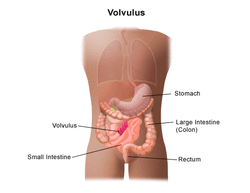
Volvulus is due to axial twisted of part of gastrointestinal tract around the mesentery. This will lead to complete or partial blocked of the bowel. Venous congestion as well as obstruction of the arterial inflow later may lead to infarction and bowel gangrenous.
There are few forms of volvulus such as gastric volvulus ( due to defect of the diaphragm ), sigmoid volvulus, transverse colon and splenic flexure volvulus and cecal volvulus as well as midgut volvulus.
Sigmoid volvulus may occur due to laxative abuse and chronic constipation. It is associated with mesocolon narrow base and redundant of sigmoid colon. Sigmoid colon mostly affect patient with psychiatric disorders,institutionalized patient, elderly and patient with chronic motility disorder of the bowel.
Cecal volvulus mostly present in pregnancy and in young adult. Cecal volvulus can be observed after colonoscopy. Cecal volvulus is associated with malabsorption /pseudo obstruction ( gas production).Cecal volvulus is due to the freely mobile cecum in varying degree as a result of improper congenital fusion of the posterior peritoneum with mesentery.
Midgut volvulus affect mostly male neonates . Midgut volvulus may present with chronic vomiting intermittently, failure to thrive, bloody diarrhea and intolerance to feeding if it affect children more than 1 years old. Due to obstruction higher in the GI tract, distention is mild in nature. Patient may have sudden onset of bilious emesis, constipation and abdominal pain . Previous episodes of bilious emesis is commonly associated with midgut volvulus. Pathologically, midgut volvulus is due to malrotation of the midgut in the utero to enter the abdomen congenitally. The whole midgut will rotate around the mesenteric stalk/ superior mesenteric artery from descending duodenum to transverse colon.
References
1.Kerry RL, and Ransom HK. “Volvulus of the Colon: Etiology, Diagnosis, and Treatment.” Archives of Surgery 99, no. 2 (August 1, 1969): 215–222. doi:10.1001/archsurg.1969.01340140087013.
2.Ghazi, A, H Shinya, and W I Wolfe. “Treatment of Volvulus of the Colon by Colonoscopy.” Annals of Surgery 183, no. 3 (March 1976): 263–265.
3.Drapanas, Theodore, and John D. Stewart. “Acute Sigmoid Volvulus: Concepts in Surgical Treatment.” The American Journal of Surgery 101, no. 1 (January 1961): 70–77. doi:10.1016/0002-9610(61)90655-9.
4.Wasselle, J A, and J Norman. “Acute Gastric Volvulus: Pathogenesis, Diagnosis, and Treatment.” The American Journal of Gastroenterology 88, no. 10 (October 1993): 1780–1784.
Patient will be admitted if suspected to suffer from volvulus. Any child who is less than 1 month old and suffer from vomiting should be suspected to suffer from volvulus. Adults are mostly not diagnose with volvulus for the first 6 months of chronic abdominal symptoms. There will be an increase in morbidity if the diagnosis is made delay especially on adult. Mortality mostly present due to gangrene.
The initial step of treatment include assessment of the airway, breathing and circulation. IV access is established. The patient is nil per os. Consider 0.9% of normal saline of 20ml/kg for children or 2 liters bolus for adult as part of the fluid resuscitation procedure. Insert the foley catheter and nasogastric tube. Surgical opinions are important and patient is prepared pre operatively . Any fluid deficit ( hypovolemia) is corrected. Electrolytes instability is also corrected. Any suspected case of perforation and sepsis are treated with broad spectrum of antibiotic pre operatively.
The treatment for specific sigmoid volvulus may include resection of the gangrenous bowel and sigmoid colon and establishing end colostomy for toxic sigmoid volvulus patient. Non toxic sigmoid volvulus is treated non operatively with reduction of volvulus non operatively via sigmoidoscopy . There will be 50% cases of recurrence which follow later with sigmoid resection electively and primary anastomosis. Cecal volvulus us treated with reductive operation and cecectomy as well as primary anastomosis. If the cecum is viable consider cecopexy.
Risk of ischemia is avoided by considering laparotomy within 1-2 hours onset. Recurrent volvulus is avoided by performing Ladd procedure, resection of the gangrenous bowel and detorsion of the bowel surgically.
Patient with volvulus may present with chronic symptoms of obstruction of the bowel. Patient may complain of crampy colicky abdominal pain, nausea and vomiting, obstipation and distended abdomen. Triad of Brochard consists of signs and symptoms such as difficulty in passing nasogastric tube, severe distention of epigastrium and intractable retching which is associated with gastric volvulus. Sigmoid volvulus usually insidious in term of onset.
Cecal volvulus may present as abdominal distention which is asymmetrical and sudden in term of onset. Cecal volvulus may also associate with sudden onset of abdominal pain.
On inspection, mass which is palpable present in the mid abdomen and left upper quadrant. This commonly associates with cecal volvulus. Signs such as tachycardia, pyrexia, tenderness, hypovolemia, peritoneal signs ( rigidity, rebound and guarding) as well as blood on digital rectal examination are common indication of gangrenous bowel.
In children, signs such as shock, abdominal pain/ distention and hematochezia are indication of necrosis/ischemia. Bilious vomiting in neonates may require surgical intervention.
Differential diagnosis of volvulus are ovarian torsion, intussusception, diverticulitis, colonic carcinoma, salpingitis, pelvic inflammatory disease, appendicitis, ileus and small bowel obstruction.
In children/neonates condition such as duodenal atresia, meconium ileus, necrotizing enterocolitis, Meckel’s diverticulum, Hirschsprung ‘s disease, appendicitis, intussusception, colic, gastroesophageal reflux, pyelonephritis, Henoch - Schonlein purpura, meningitis and inborn error of metabolism are associated with volvulus.
It is extremely important to assess any children with evidence of obstruction ( abdominal pain and bilious vomiting) for malrotation as any late diagnosis may require permanent paraenteral nutrition and large resection due to gangrenous bowel.
The investigations required are full blood count, urea and electrolytes, urinalysis, abdominal radiography, barium enema, CT scan and ultrasound scan. Full blood count may reveal leukocytosis more than 20 000 in cases of peritonitis and strangulation. Urea and electrolytes studies reveal dehydration due to pre renal azotemia and anion gap acidosis due to lactic acidosis. Urinalysis may reveal the present of ketones and elevated specific gravity.
Cecal volvulus may present with dilated and displaced loop of small bowel with kidney shaped cecum in the left abdomen. Abdominal radiography may reveal U shaped inverted loop of dilated colon from the pelvis.
Barium enema may have a therapeutic purpose. The procedure should be done carefully to avoid perforation and may reveal bird beak at the site of torsion. Series of upper GI imaging may reveal corkscrew tapering of the contrast and abrupt ending. CT scan is useful in identifying the extent obstruction in case of sigmoid volvulus. In cecal volvulus whirl sign may present. Ultrasound may also reveal whirlpool signs of volvulus where the vessel twirled around the mesentery base. Ultrasound may reveal abnormal position of superior mesenteric vein.
Patient with sigmoid volvulus will get the benefit from endoscopic decompression with placement of rectal tube. However recurrence is common. Elective surgical treatment may be required.
Volvulus is due to axial twisted of part of gastrointestinal tract around the mesentery. This will lead to complete or partial blocked of the bowel. Venous congestion as well as obstruction of the arterial inflow later may lead to infarction and bowel gangrenous.
There are few forms of volvulus such as gastric volvulus ( due to defect of the diaphragm ), sigmoid volvulus, transverse colon and splenic flexure volvulus and cecal volvulus as well as midgut volvulus.
Sigmoid volvulus may occur due to laxative abuse and chronic constipation. It is associated with mesocolon narrow base and redundant of sigmoid colon. Sigmoid colon mostly affect patient with psychiatric disorders,institutionalized patient, elderly and patient with chronic motility disorder of the bowel.
Cecal volvulus mostly present in pregnancy and in young adult. Cecal volvulus can be observed after colonoscopy. Cecal volvulus is associated with malabsorption /pseudo obstruction ( gas production).Cecal volvulus is due to the freely mobile cecum in varying degree as a result of improper congenital fusion of the posterior peritoneum with mesentery.
Midgut volvulus affect mostly male neonates . Midgut volvulus may present with chronic vomiting intermittently, failure to thrive, bloody diarrhea and intolerance to feeding if it affect children more than 1 years old. Due to obstruction higher in the GI tract, distention is mild in nature. Patient may have sudden onset of bilious emesis, constipation and abdominal pain . Previous episodes of bilious emesis is commonly associated with midgut volvulus. Pathologically, midgut volvulus is due to malrotation of the midgut in the utero to enter the abdomen congenitally. The whole midgut will rotate around the mesenteric stalk/ superior mesenteric artery from descending duodenum to transverse colon.
References
1.Kerry RL, and Ransom HK. “Volvulus of the Colon: Etiology, Diagnosis, and Treatment.” Archives of Surgery 99, no. 2 (August 1, 1969): 215–222. doi:10.1001/archsurg.1969.01340140087013.
2.Ghazi, A, H Shinya, and W I Wolfe. “Treatment of Volvulus of the Colon by Colonoscopy.” Annals of Surgery 183, no. 3 (March 1976): 263–265.
3.Drapanas, Theodore, and John D. Stewart. “Acute Sigmoid Volvulus: Concepts in Surgical Treatment.” The American Journal of Surgery 101, no. 1 (January 1961): 70–77. doi:10.1016/0002-9610(61)90655-9.
4.Wasselle, J A, and J Norman. “Acute Gastric Volvulus: Pathogenesis, Diagnosis, and Treatment.” The American Journal of Gastroenterology 88, no. 10 (October 1993): 1780–1784.
