Medicine Notes - Clinical Procedures - Chest Drain Insertion

Chest drain insertion
The insertion of chest drain may involve the use of Seldinger technique. The indication of chest drain insertion may include to relief pleural effusion, chylothorax, hydrothorax, empyema, pneumothorax and hemopneumothorax.
The equipments require for the insertion of chest drain include 25 G needle, 21G needle, 10ml syringe, 10ml 10% lidocaine, suture, sterile pack, sterile gloves, cleaning solution, dressing, chest drain, drain kit needle, syringe, guidewire,3 way tap and scalpel.
The procedure is avoided /contraindicated if patient develop skin infection at the site of insertion, large bullae, pleural adhesion, thoracic adhesion, coagulopathy and cases of emergency thoracotomy.
The position of the patient is important. Patient need to be sitting on a chair or on the edge of the bed.
Clinical examination and chest radiography are performed to reassure the sides which need to be drained.
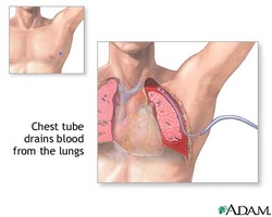
The chest drain need to be inserted in “ triangle of safety”. The chest drain is inserted within a triangular area formed by pectoralis major, diaphragm and latissimus dorsi. The site is usually at the mid axillary line above the rib to avoid hitting the neurovascular bundle.
Sterile gloves are put. The area of insertion need to be cleaned with antiseptic solution / cleaning solution ( iodine/ chlorhexidine).
The 10 ml syringe and 25 G needle/ orange needle are used to anesthetize the skin by creating a subcutaneous bleb.
The 21G needle / green needle is used to anesthetize the pleura.
A scalpel is used to make small incision of the skin.
A syringe , curved tip and drain kit needle are used to advance through the anesthetized area with curve tip facing downwards. It is advanced until air and fluid are aspirated.
The needle is hold in place while the syringe is removed.
The guidewire is threaded through the needle and later into the chest. Then the covering is removed.
The needle is withdrawn from the chest but guidewire should remain there.
The introducer is threaded over the guidewire and twisted it back and forth to open a tract to drain the passage. The introducer then need to be slide back off.
The drain is then threaded over the wire and into the chest. Curving downward with the central stiffener in place.
The stiffener and wire are removed once the drain is in place. The 3 way taps are attached later. Drain is stitches in place.
The drain is attache to the tubing. The tubbing is attached to the collecting bottle with 500 ml sterile water.
The 3 way taps need to be open. Fluid or air start to flow in the collecting bottle. Patient may take a few breath and the water level in the tubing will be swinging ( rise and fall).
Chest radiography is performed to confirm the position of chest drain insertion.
The procedure carries its own risks which include local bleeding, iatrogenic placement, hemothorax, infection, penetration to organs ( heart, diaphragm, colon, liver, stomach, spleen,lung) and injury to the liver or spleen, hemoperitoneum and inadequate placement.
