|
|
|
Emergency Strategy - How to treat fracture of the femur

Emergency strategy - How to treat fracture of the femur
The initial treatment of fracture of the femur include the airway, breathing , circulation, disability and exposure stabilization. Blood pressure is monitored and treated as hypotension may present as a result of hemorrhagic shock with potential loss of 4- 6 units of blood. The extremity is immobilized and traction split is applied for tamponade of further loss of blood into the thigh.
The extremity is support in a position of comfort by backboard immobilization and rigid splinting. Traction is avoided in case of fracture of the leg, fracture of the pelvis, fracture of the knee and dislocation or fracture of the ipsilateral hip.
Sterile dressing is used to cover the open wound.Do not try to reduce the open fracture. Any airway, abdominal and chest injuries should be observed and treated if present.
The lower extremity should be maintained stable. Pain control is prescribed to the patient. Consider parenteral analgesic in case of isolated injuries to the femur. Femoral nerve block is considered in case of pediatric setting or multi traumatic patient. Orthopedic consultation is important. Debridement and irrigation are considered in case of open fracture in operation theatre. Skeletal traction will be considered in case the patient is not immediately going to the operation theatre. Femoral artery exploration and angiography are considered if the femur fracture is associated with palpable pulsatile mass ( aneurysm), hematoma expanding and absent of distal pulses. Neurovascular compromise required an emergent /urgent treatment.
Antibiotic such as cefazolin is considered in case of open fracture of the femur with laceration, contamination, extensive damage to the tissue. Tetanus injection is also considered as well as additional tobramycin /gentamicin. Penicillin G is considered in highly contaminated wound which cover the species of clostridia.
Patient with fracture of the femur need to be admitted and patient will later discharge with pain control medication. Patient with pathological fracture or would not undergo operative fixation and not ambulatory may be discharge with pain control and agreement from the orthopedic.
Patient with fracture of the femur may present with inability to move the knee and hips. They may complain of pain in the thigh and shortening, swelling and deformity of the thigh. There is also evidence of multi injuries such as airway, abdominal and chest injuries. Hypotensive patient is common and associate with hemorrhagic shock due to hemorrhage into the thigh. Patient may suffer from penetrating trauma which leads to open fracture. Compartment syndrome /vascular compromise may occur due to impair circulation of the food. It is important to assess the neurovascular status. The common complication from the fracture of the femur include compartment syndrome, hemorrhage, adult respiratory syndrome and fat embolism.
The fracture of the femur is often mistaken with hematoma, dislocation of the hip, fracture of the hip, contusion of the thigh and dislocation of the knee and fracture of the knee.
The investigations require are full blood count, type and cross match of the blood. Angiography and doppler are considered if the pulses are absent.
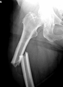
Radiography such as AP pelvis, AP and lateral view of the femur, true lateral of the hip and complete series of radiography of the knee are considered as well as chest x ray, Bone scan and skeletal survey should be oredered in case of suspected non accidental trauma in pediatric setting.
Fracture of the femur can be classified based on the geometry, location, extent of injury to the soft tissue and degree of comminution. Geometrically fracture of the femur can be divided into spiral fracture of the femur, transverse fracture of the femur, oblique fracture of the femur and segmental fracture of the femur. Fractures of the femur can be divided into proximal third of the femur which involve the subtrochanteric region, middle third and distal third of the femur which involve the distal metaphyseal - diaphyseal junction. Femur fracture can also be divided into open fracture of the femur and closed fracture of the femur based on the degree of damage to the tissue.
Winquist and Hansen classification is useful in identifying the degree of comminution. Grade 1 fracture of the femur involve small fragment less than 25% width of the femoral shaft. The femur is rotationally stable with stable lengthwise. Grade 2 fracture of the femur involves 25 - 50 % width of the femoral shaft . The femur may /may not loss its rotation stability and it still stable lengthwise. Grade 3 fracture is unstable lengthwise and rotationally. The fracture involves more than 50% of the width of the femoral shaft, Grade 4 fracture of the femur is unstable lengthwise and rotationally and lead to loss of cortex circumferentially.
Fracture of the femur is usually caused by high energy injury such as trauma due to fall ( spiral fracture), gunshot wounds and major traffic accident. Repetition activity such as heavy exercise, work load may also lead to stress fracture which contributes to fracture of the femur. Pathological fracture may also lead to fracture of the femur.
In pediatric setting, fracture of the femur may be associated with non accidental trauma. Child with fracture of femur may present with bruises, burns, linear abrasion and isolated trauma to the thigh. There will be a delay in presentation and dislocation of the femoral capital epiphysis may present. Spiral fracture of the femur in children is an indication of non accidental trauma. Non accidental trauma account for 70% of femoral fracture in children less than 3 years old.
References
1.Anderson, William A. “The Significance of Femoral Fractures in Children.” Annals of Emergency Medicine 11, no. 4 (April 1982): 174–177. doi:10.1016/S0196-0644(82)80492-7.
2.Evans, E. Mervyn. “The Treatment of Trochanteric Fractures of the Femur.” Journal of Bone & Joint Surgery, British Volume 31-B, no. 2 (May 1, 1949): 190–203.
3.Reeves, R B, R I Ballard, and J L Hughes. “Internal Fixation Versus Traction and Casting of Adolescent Femoral Shaft Fractures.” Journal of Pediatric Orthopedics 10, no. 5 (October 1990): 592–595.
