Symptom finder - The causes of urinary incontinence

Symptom finder - The causes of urinary incontinence
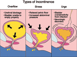
The causes of urinary incontinence are anatomical such as vesicovaginal fistula, ectopic ureter or ectopia vesicae , stress incontinence such as pelvic floor injury due to prostatectomy or childbirth, neurological causes such as upper motor neuron lesion due to syringomyelia , multiple sclerosis, spinal tumors, spinal cord injury above the sacral center, head injury, cerebrovascular accident and disc lesion, overflow incontinence due to chronic outflow obstruction such as prostatic enlargement or lower motor neuron lesions due to damage to sacral nerve form pelvic surgery, cauda equina injury, sacral center injury and diabetes and finally urinary incontinence may be due to urge incontinence such as detrusor instability due to tumor, interstitial nephritis, tuberculo cystitis , post radiotherapy, prostatectomy, stones and cystitis. Nocturnal enuresis may also cause urinary incontinence.
What is urinary incontinence ? Urinary incontinence is the loss of urine involuntary from the bladder which is inconvenient, socially embarrasing, inappropriate and occur at any times or places. Urge incontinence is frequent and persistent urge to void and the individual is unable to maintain the urinary incontinence. Stress incontinence is the lose of urine during sneezing, coughing and straining. Distended bladder due to flaccidity of the detrusor muscle which become insensitive to stretch may lead to overflow incontinence. The sphincter mechanism is weak and later leads to overflow as the urine leak from the urethra.
Incontinence with saddle anesthesia and weakness of the leg suggestive of cauda equina lesion which require urgent referral to neurosurgical unit. Continuous incontinence suggestive of neurological causes, chronic outflow problems and fistula.
Anatomically erosion of the ureteric calculus into fornix of the vagina may lead to ureterovaginal fistula. Pelvic radiotherapy or pelvic surgery may lead to vesicovaginal fistula. which is the presenting symptoms of pelvic malignancy. Ectopia vesicae will present at birth with ureter opening into bladder mucosa which is exposed on the lower abdominal wall as well as defect in an abdominal wall.
Patient with history of loss of urine during sneezing and coughing , recent prostatectomy, difficulty in child delivery or multiple birth are predispose to stress incontinence.
Patient with history of syringomyelia, multiple sclerosis, cerebrovascular accident, head injury and trauma to the spinal cord affecting the cord above sacral centre may present with upper motor neuron lesion which predispose to neurological causes of urinary incontinence.
Overflow incontinence is associated with diabetes, abdominoperinal resection of rectum ( pelvic surgery), damage to pelvic nerve and prostatism with retention and overflow which is chronic in nature. Patient with overflow incontinence may be able to void normally but he may feel the leakage of the urine is continue and the bladder is not emptying enough.

Urge incontinence mostly present in patient with hematuria due to tumor or stones, persistent discomfort of the suprapubic region, sufferer of ureteric colic, TB patient , previous history of pelvic radiotherapy, recent prostatectomy and dysuria or frequency due to recurrent attack of cystitis. 5% of children ( 10 years ) may present with nocturnal enuresis . After puberty, the present of bed wetting indicates the present of pathological condition of the bladder.
On examination, ectopia vesicae is visualized as ureters that open into mucosa of the bladder on the abdominal wall. Vaginal speculum examination may show sites/location of the fistula. Full neurological examination is perfored. Reflex emptying of bladder does occur. In stress incontinence a complete prolapse or cystocele may be seen. Patient is observed while coughing to look for any leakage.
In overflow incontinence, the bladder may be palpable. Prostatic hypertrophy is detected with digital rectal examination. Full neurological examination is performed.
Laboratory investigation may include full blood count, mid stream urine, intravenous urography, cystography, cystoscopy, CT scan / MRI scan and urodynamic.
Full blood count may reveal raised white cell count due to infection. Mid stream urine is useful for culture and sensitivity to detect any infection. Intravenous urography is useful for observing the upper urinary tract for any fistula, lesion on the bladder and obstruction. Cystography is useful for detection of vesicovaginal fistula. Neoplasia, or stone in the bladder is detected with cystoscopy. Urodynamics studies may include urethral pressure profile to assess outlet obstruction and sphincter function, videocystometry for any leakage of urine while the patient is straining in stress incontinence. Detrusor contraction is assessed with cystometry and flow rates is measured with uroflowmetry.
