|
|
Pathology definition - Malignant Melanoma
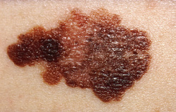
Malignant melanoma
Malignant melanoma may present with variation in terms of border, sizes and pigmentation. There are few variation of malignant melanoma. The first variant of malignant melanoma is superficial spreading. Superficial spreading of the malignant melanoma is the commonest form of malignant melanoma which present with irregular border. There is no invasion of the dermis and superficial spreading malignant melanoma undergoes radial phase of growth ( malignant melanoma which grow horizontally over the skin with no deep invasion). The second variant is the nodular form of malignant melanoma which undergoes vertical growth phase and nodular in nature. The third variant of malignant melanoma is known as lentigo maligna. Lentigo maligna arises from the Hutchinson freckle which is a precursor lesions . lentigo maligna later will give rise to desmoplastic melanoma or spindle cell melanoma.
Malignant melanoma occurs as a result of fair skin, genetic predisposition and excessive exposure to the sunlight as well as patient who is immunosuppressive. The treatment of malignant melanoma may focus on the chemotherapy, dissection of the regional lymph node ( if involved) and the main treatment ( surgical excision).
Malignant melanoma usually arises from melanocytes and nevus cell. The cluster of nevus cells will demonstrate anaplasia and cellular atypia. The cluster of the nevus cell usually present within the epidermis. There will be linear fibrosis and mild infiltration of the lymphocytes. This is known as dysplastic nevus. There a high chances that the dysplastic nevus may progress into malignant melanoma. Dysplastic nevus is known as precursor lesions. Malignant melanoma is detected by the present of tumor marker known as S - 100
Malignant melanoma may undergo two form of growth phase patterns. These includes vertical growth and radial growth. Vertical growth is a growth into the dermis. Vertical growth usually involves the nodular pattern of the malignant melanoma which carries a poor prognosis. Vertical growth malignant melanoma able to metastasis (eg. to the meninges, eyes, esophagus) based on the depth of the invasion. Radial growth phase does not metastasis. Radial growth phase will growth horizontally and consists of cluster of atypical cells within the epidermis. Radial growth malignant melanoma consists of discolored macule with the present of melanin containing macrophages in the dermis and infiltration of the lymphocytes.
References
1.McGovern, Vincent J., Martin C. Mihm, Christiane Bailly, Joan C. Booth, Wallace H. Clark, Alistair J. Cochran, E. G. Hardy, et al. “The Classification of Malignant Melanoma and Its Histologic Reporting.” Cancer 32, no. 6 (1973): 1446–1457. doi:10.1002/1097-0142(197312)32:6<1446::AID-CNCR2820320623>3.0.CO;2-8.
2.Harpio, Riikka, and Roland Einarsson. “S100 Proteins as Cancer Biomarkers with Focus on S100B in Malignant Melanoma.” Clinical Biochemistry 37, no. 7 (July 2004): 512–518. doi:10.1016/j.clinbiochem.2004.05.012.
