Symptom finder - The causes of swelling of the leg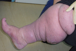
Symptom finder - The causes of swelling of the leg.
Swelling of the leg or lower limb can be divided into unilateral, bilateral, localized and general swelling of the leg. Trauma, lymphatic disorders and venous disorders are common causes of unilateral/one sides swelling of the leg. Renal failure, cardiac failure and hepatic failure are common medical condition which lead to bilateral/both sides swelling of the leg. Generalized swelling of the leg are commonly caused by renal failure, myxoedema, congestive cardiac failure , overload of fluid and hypoproteinemia ( malnutrition, nephrotic syndrome and liver failure). Localized swelling is categorized into chronic causes of swelling and acute cases of swelling . Chronic cases of swelling are due to arteriovenous fistula dependency, failure of muscle pump, lymphedema ( unilateral, non pitting edema of the young)and venous causes. Patient may have underlying malignancy in cases of deep vein thrombosis related swelling. Other causes of chronic swelling include post - phlebitic limb, inferior vena cava obstruction, tumor of the pelvic and pregnancy. All of these are causing obstruction to the venous return. Trauma, allergy, rheumatoid arthritis, cellulitis and deep vein thrombosis account for acute swelling of the leg. |
|
History will focus on the sites of the swelling, the onset of the swelling , the predisposing factors for swelling and associated symptoms. Associated symptoms are mostly painful and painless. Painful leg swelling are due to infection, deep vein thrombosis and varicose vein. Painless but uncomfortable swelling is due to tense edema.
The history will focus on past medical history. These includes malnutrition, nerve lesions, liver failure, poliomyelitis, renal failure, thyroid disease, previous surgery, plevic or abdominal malignancy, radiotherapy to the lymph nodes, congestive cardiac failure, limb’s trauma and pregnancy.
Primary lymphedema present as swollen/edematous leg since birth. lymphedema precox occur at puberty ( edema of puberty) and lymphedema tarda is an edema that occur in the third decades.
Physical examination will focus on inspection. Rheumatoid arthritis is suggestive by tenderness and swelling over the joint. Positive Homan’s sign ( calf pain while dorsiflexing the foot), tender and swollen limb are highly suggestive of deep vein thrombosis. Lymphedema is suggestive by a mass of malignant nodes, scar in the groin and previous radiotherapy. In early stages, lymphedema pits. In later stages as the subcutaneous and skin become warty, hyperkeratotica and thick the lymphedema become non pitting. Inferior vena cave thrombosis is suggested by dilated cerebral vein of the wall of the abdomen and bilateral swelling. Femoral vein thrombosis may present with tenderness along the femoral vein. Phlegmasia alba dolens or swollen pale limb to the groin or phlegmasia cerulea dolens or purplish, tense and painful limb are associated with iliofemoral thrombosis. Any contusion, fracture or hematoma will be obvious . Look for any visible puncture wound and consider cellulitis in case of swollen, red hot as well as tender limb.
Tumor may present as localized swelling of the leg. Distal swelling may occur as a result of lymphatic and venous obstruction. Dilated vein which do does not collapse while the limb is elevated is a characteristic of arteriovenous fistula. Other characteristic of arteriovenous fistula are continuous machinery bruit on auscultation and thrill on palpitation.The limb with arteriovenous fistula will appear warmer than opposites. Any mass on the abdomen or hepatomegaly may reveal the venous outflow obstruction. Pelvic tumor will be detected using rectal examination. The pelvic tumor is characterized as back pressure against the lymphatic system and venous system that result in frozen pelvis. Swelling as well as wasting may occur due to nerve lesions or poliomyelitis.
The investigation require are full blood count, urea and electrolytes, ESR, urinalysis, creatinine, liver function test, chest x ray, limb x ray, blood glucose, clotting screen, lymph node biopsy, lymphangiography, arteriography, venography, Duplex doppler, CT scan, thyroid function test, ultrasound scan, MRI scan and ultrasound or CT scan of the pelvis.
Full blood count may reveal low Hb level which is associated with fracture or trauma due to large hematoma. Reduction in platelet count may also occur due to hematoma. Increase in white cell count is an indication of infection. Urea and electrolytes studies may reveal raised urea and creatinine which is an indication do chronic renal failure. Proteinuria in urinalysis is suggestive of renal failure. Liver function test may reveal the present of hypoalbuminemia that indicates an impairment of liver function.
Chest x ray may reveal secondary deposits due to sarcoma of the limb. Chest x ray may also detect the signs of cardiac failure such as pleural effusion, pulmonary edema ( secondary to fluid overload due to renal failure ) and cardiomegaly.
X ray of the limb may reveal the gas in tissue ( gas gangrene), tumor or fracture. Diabetes is detected with measurement of the blood glucose level. Diabetes may leads to infections and cellulitis.
Clotting screen may reveal abnormality in blood clotting due to coagulopathy from spontaneous hematoma. Biopsy of the lymph node is vital to exclude any tumor or infection. The causes of lymphedema such as obstruction or hypoplasia are detected by using lymphangiography. Arteriovenous fistula is detected by arteriography.Deep vein thrombosis is detected through venography. Duplex doppler is useful to detect arteriovenous fistual and deep vein thrombosis. Ultrasound is useful to rule out soft tissue sarcoma or hematoma. Ultrasound and CT scan of the pelvis may reveal the present of abdominal mass that exert extrinsic pressure to the veins. MRI scan is useful for detection of soft tissue sarcoma. CT scan is useful for detection of hematoma or tumor. Thyroid function test is useful to rule out myxoedema.
