
Emergency Strategy - How to treat atrioventricular heart block
 ECG of heart trace
Emergency Strategy - How to treat atrioventricular block
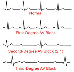
There are three different forms of atrioventricular block which include first degree AV block, second degree AV block and third degree AV block. First degree AV block causes include myocarditis, toxicity of digoxin, increase vagal tone and acute inferior myocardial infarction. First degree AV block is characterized by delay in AV conduction. In certain cases, first degree AV block, may involve normal heart. None is required in term of management as the patient is mostly asymptomatic. However, ECG may show prolongation of the PR interval more than 0.20 seconds.
Second degree AV block is characterized by intermittent AV conduction. Some of the impulses from the atrium reach the ventricles while some are blocked. There are two different forms of second degree AV block such as second degree Mobitz type 1 /Wenckebach AV block or second degree Mobitz type 2 AV block.
Mobitz type 1 second degree AV block is characterized by transient reversible depression of the AV nodes of conduction. Mobitz type 1 AV block usually involve the level of thee AV nodes. ECG of the patient with Mobitz type 1 may reveal progressively prolongation of the PR interval before the blockage of the conduction of the atrium impulses. Prior to the blocked P waves , the last conducted impulse from the atrium appear to be longer than the PR interval of the first impulses conducted. Myocarditis, toxicity of the digoxin and acute inferior myocardial infarction may causes second degree Mobitz type 1 / Wenckebach AV block. Treatment with 0.5 mg of IV atropine is given and every 5 minutes the dose is repeated until it achieves the total dose of 2 mg especially in specific case that may lead to hypoperfusion.
Mobitz type 2 AV block / high grade block is associated with damage structurally to the infranodal conducting system. Mobitz type 2 AV block may be permanent and later progress to complete heart block. ECG may reveal sudden and unexpected fail in conduction without any changes in the PR interval. ECG may also reveal prolongation of the PR interval. Mobitz type 2 may present with high grade block or constant AV block ration eg. 3:! means P waves 3 times more frequent than the QRS complex. In term of management, permanent transvenous cardiac pacing will be inserted in most cases. Hypoperfusion is an indication for the use of isoprenaline and atropine . However, isoprenaline is hazardous to digitalis toxicity or acute myocardial infarction.
Third degree AV block is also known as complete heart block. Generally, ECG may reveal completely dissociated regular P waves from QRS complex . In certain cases, QRS is widened or normal with variation of the PR interval.Third degree AV block will lead to disruption of the AV conduction. In certain cases, there is no AV conduction . Pacing of the ventricles occur due to escape pacemaker at a rate shorter than the atrial rate.
Third degree AV block is further divided into nodal block and infranodal block. Nodal block occurs as a result of depression of the conduction and carries no serious prognosis. Nodal block usually occur as a result of congenital block or inferior myocardial infarction . Nodal block may present with ventricular rates of 40 - 60 bpm and narrow QRS complex. The management of nodal third degree heart block is similar to Mobitz type 1 AV block.
Infranodal third degree heart block carries a serious prognosis with unstable and slow ventricular escape rhythm. Infranodal third degree heart block mostly occur due to organic disease ( idiopathic fibrosis and acute myocardial infarction) which affect the bundle of His or bundle branches. The ECG may reveal ventricular rates of less than 40 bpm with widened QRS complex . Patient with infranodal third degree AV block may require pacemaker. Ventricular escape rhythm is temporary accelerated with isoprenaline.
References
1.Mymin, David, Francis A.L. Mathewson, Robert B. Tate, and Jure Manfreda. “The Natural History of Primary First-Degree Atrioventricular Heart Block.” New England Journal of Medicine 315, no. 19 (1986): 1183–1187. doi:10.1056/NEJM198611063151902.
2.Benedict, Ruth B., and John M. Evans. “Second-degree Heart Block and Wenckebach Phenomenon Associated with Anxiety.” American Heart Journal 43, no. 4 (April 1952): 626–633. doi:10.1016/0002-8703(52)90124-5.
3.Huhta, J. C., J. D. Maloney, D. G. Ritter, D. M. Ilstrup, and R. H. Feldt. “Complete Atrioventricular Block in Patients with Atrioventricular Discordance.” Circulation 67, no. 6 (June 1, 1983): 1374–1377. doi:10.1161/01.CIR.67.6.1374.
4.Burton, C. R., Michael M. Abbott, and Kenneth Mitsui. “Heart Block in Adult Patients in Hospital Practice.” Canadian Medical Association Journal 84, no. 9 (March 4, 1961): 461–465.
5.Langendorf, Richard, and Alfred Pick. “Atrioventricular Block, Type II (Mobitz)—Its Nature and Clinical Significance.” Circulation 38, no. 5 (November 1, 1968): 819–821. doi:10.1161/01.CIR.38.5.819.
