Emergency Strategy - How to treat pemphigus
|
|
|

Emergency Strategy - How to treat pemphigus
There are a few skin disorders that share the similar symptoms and signs such as pemphigus. These includes bullous pemphigoid, toxic epidermal necrolysis, eryhroderma, erythema multiforme, epidermolysis bullosa, contact dermatitis, hand, foot and mouth disease, dermatitis herpetiformis, erysipelas, seborrheic dermatitis, oral candidiasis, systemic lupus erythematosus, systemic vasculitis, lichen planus and herpes simplex gingivostomatitis.
The diagnosis of pemphigus is confirmed based on the history of the presenting complain, clinical assessment and laboratory investigations.
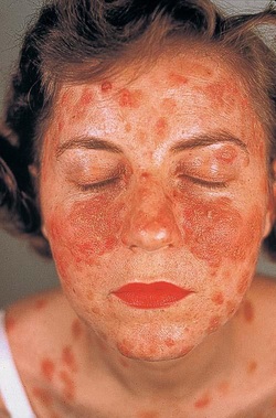
Patient may present with non - healing and painful erosion of the mucosal, vaginal or oral region. The mucocutaneous blister will initially present which later develop into erosion. It may affect the mucous membrane initially before progressing into skin/ cutaneous layer. The blister usually present as localized or generalized lesion. Erosion of the skin may lead to detachment of the epithelium. Nikolsky sign may present ( separation of the epidermis from lateral pressure. The bullae may be rupture and later present as crusting partially healed skin. Pemphigus may also present as edematous, exfoliate, moist erosion in seborrheic areas or hyperplastic/hypertrophic erosive plague with pustule in intertriginous area. Pemphigus also may be distributed in malar distribution as scaly, crusty, erythematous lesion of the skin.
The investigation require are full blood count, urea and electrolytes, serum antibody titers, immunofluorescent studies and biopsy of the skin.
In acute setting, after assessment and stabilization of the patient’s airway, breathing and circulation consider monitoring the vital sign, cardiac monitoring and establish IV lines for fluid resuscitation in case of sepsis or severe hypotension based on the Parkland formula. In this formula, 4ml/kg of crystalloid solution is given for 24 hour based on the percentage of the body surface involvement. Half of the fluid is given for the first 8 hours and the following half for the next 16 hours. Urine output should be maintained to be more than 0.5ml/kg/hr.
Steroid is considered in the treatment of pemphigus. Stress dose of steroid is given. In severe cases, high dose of corticosteroids are administered. Pulse IV corticosteroids is considered if the patient is unresponsive to treatment with oral corticosteroids. Patient may also be admitted for plasmapheresis. Floor bed is usually consider while treating the patient with plasmapheresis or pulse parenteral steroid therapy.
Vasopressor is given to patient to keep the mean arterial pressure more than 65mmHg, central venous pressure 8 - 12 mmHg and central venous oxygen saturation more than 70%. Dapsone , IV immunoglobulins, cyclophosphamide, azathioprine, gold, mycophenolate, methotrexate and cyclosporin are immunosuppressive therapy which is given to reduce the side effect of high dose of corticosteroids. The immunosuppressive therapy may lead to hyperosmolar non ketotic acidosis, type 2 diabetes mellitus,sepsis and adrenal crisis.
Intralesional triamcinolone acetonide may also be given to the patient. If the patient develop sepsis, broad spectrum antibiotic is presented.
Patient is admitted to the burn units or ICU if sepsis is suspected and the patient will develop shock. Patient will be discharged if the condition is mild and moderate and a follow up with dermatologist is required. Patient with high dose of steroid may develop osteoporosis. In this case bone scan is performed and patient is referred to rheumatologist as part of the follow up.
How pemphigus develop? Generally there are three different form of pemphigus. Pemphigus foliaceus ( erosion which is edematous , exfoliate and moist in seborrheic area), pemphigus vegetans ( erosive plaque and pustule which is hypertrophic and hyperplastic in nature) and pemphigus erythematosus ( scaly crusty, erythematous lesions of the skin in malar distribution. Pemphigus develop due to action of IgG autoantibodies against desmosomal cadherins desmoglein 1 and 3 which present in all keratinocytes. This will result in acantholysis or loss of adhesion between cells. Acantholysis will be worse due to exposure to UV B from the sunlight. Loss of cell - cell adhesions/acantholysis and separation of keratinocytes may lead to formation of bullae.
Pemphigus foliaceus usually mild and superficial. Pemphigus vulgaris usually serious and involved deep structures. Most commonly present as oral lesions. Mortality rates is highest in case of mucocutaneous involvement.
Pemphigus is associated with lymphoma ( paraneoplastic pemphigus). Drugs may also lead to pemphigus such as rifampicin, penicillamine and phenobarbital. Patient with specific human leukocyte antigen HLA haplotypes ( DRW6 and DR4 ) are also predispose to pemphigus. Any bites from flying insect may also lead to pemphigus foliaceus which is an endemic in south american.
Pemphigus is a rare disorders. It is common in individual age 40 -60 years old. It is rare in neonates and occur due to transplacental transfer of IgG. It will resolved spontaneously as the maternal autoantibodies catabolized. It affects male and female equally.
References
1.Harman, K.e., S. Albert, and M.m. Black. “Guidelines for the Management of Pemphigus Vulgaris.” British Journal of Dermatology 149, no. 5 (2003): 926–937. doi:10.1111/j.1365-2133.2003.05665.x.
2.Ruocco, V., A. Rossi, G. Argenziano, C. Astarita, L. Alviggi, B. Farzati, and G. Papaleo. “Pathogenicity of the Intercellular Antibodies of Pemphigus and Their Periodic Removal from the Circulation by Plasmapheresis.” British Journal of Dermatology 98, no. 2 (1978): 237–241. doi:10.1111/j.1365-2133.1978.tb01630.x.
3.Becker, B A, and A A Gaspari. “Pemphigus Vulgaris and Vegetans.” Dermatologic Clinics 11, no. 3 (July 1993): 429–452.
